
Dental computed tomography (CT) scans are a relatively new technology used by dental professionals to get 3D images of the teeth, jaws, and surrounding structures. This technology is particularly useful in surgical planning for dental implants, as it can provide detailed information on bone quality and nerve canals.
CT scans use cone beam CT (CBCT) to create highly accurate images that allow dentists to assess the extent of a patient’s dental care needs. The process involves sending X-rays through the area of interest and capturing the resulting data, using a computerized system. This data is then used to construct an image in three dimensions. Dentists can use these images to identify potential problems such as impacted teeth, cysts or tumors, bone defects, or nerve canal involvement that could affect treatment.
In addition to being beneficial for implant placement, dental CT scans can also help with diagnostics and treatment planning for other procedures such as root canal therapy or wisdom tooth extractions. They can also be used to check for areas of decay around existing fillings or crowns, as well as to evaluate orthodontic treatments like braces or Invisalign.
Overall, dental CT scans are an invaluable tool for periodontists and other dentists who need more detailed information than traditional radiographs provide in order to properly plan and provide effective treatment. By providing valuable information about bone structure, nerve canals, and other relevant details within seconds rather than minutes or hours, they allow dental professionals to make fast and informed decisions about their patients’ care.
Advantages of Dental CT Scans
Accurate Diagnosis and Treatment Planning for Patients
Due to their ability to capture detailed 3D images of dentofacial structures, dental CT scans are extremely helpful in making a proper diagnosis and treatment plan for patients. The scans provide detailed information about potential dental issues, which can help dentists accurately diagnose and develop more effective treatment plans. This is especially beneficial for complex cases involving implants, root canals, wisdom teeth extractions, and orthodontic treatments such as braces or Invisalign.
The accuracy of the scans also allows dentists to identify areas of decay around existing fillings or crowns that might not be visible with traditional radiographs. This helps to ensure that any potential problems are addressed before they worsen and cause further damage. Furthermore, the 3D images produced by the CT scan allow dentists to view the mouth from all angles in order to get an accurate picture of what is going on inside the mouth. This helps them to assess any potential risks associated with various dental treatments and make informed decisions accordingly.
Overall, dental CT scans significantly improve accuracy in diagnosis and treatment planning for patients by providing detailed information about their oral health quickly and easily. By using this technology, periodontists are able to accurately identify potential problems that might otherwise go unnoticed until it is too late to properly address them. As a result, this ensures that patients receive proper care with minimal risk of complications or negative outcomes due to misdiagnosis or incorrect treatment planning.
More Detailed Images than Traditional X-Rays
Dental CT scans provide a more detailed and accurate view of the teeth and surrounding structures than traditional dental X-rays. Unlike traditional X-ray films, which produce two-dimensional images, the 3D scans created by dental CTs provide a clearer picture that allows dentists to easily identify potential problems such as impacted teeth, cysts or tumors, or bone defects. Furthermore, the higher-quality images produced by the scan also allow for more precise tooth orientation, which helps with implant placement and other delicate procedures. Additionally, these images can be viewed from any angle in order to better assess any potential risks associated with various treatments.
Overall, dental CT scans are a valuable tool for dentists because they provide more detailed images than traditional X-rays. These highly accurate 3D scans allow dentists to diagnose and develop effective treatment plans for their patients quickly and easily. As a result, this ensures that patients receive proper care with minimal risk of complications or negative outcomes due to misdiagnosis or incorrect treatment planning.
Minimal Radiation Exposure for Patients
One of the main advantages of using dental CT scans over traditional X-rays is that they result in minimal radiation exposure for patients. This is because the X-ray source used to produce these scans is significantly more powerful than the one used in traditional dental practices. As a result, it requires less time to capture 3D images, which allows for shorter scanning sessions and, thus, lower doses of radiation. In addition, because the scan captures a much larger field of view compared to traditional X-ray films, dentists are able to produce highly detailed images with fewer exposures. This minimizes radiation exposure even further and ensures that patients receive proper care with minimal risk of any complications due to excessive radiation exposure. All in all, dental CT scans provide an excellent way to receive accurate and detailed information about oral health with minimal risk of radiation exposure for patients.
Ability to Visualize Soft Tissues, Bone Structure, and Nasal Cavities
Dental CT scans provide a number of advantages in terms of accurately visualizing soft tissue structures, bone structure, and nasal cavities. The ability to view these areas in such detail is incredibly useful for periodontists when performing procedures such as dental implants and other related treatments. The increased accuracy of the scan allows dentists to place implants with greater precision than ever before, helping to reduce potential complications and risks associated with the procedure. Additionally, the highly detailed images produced by the scan also allow dentists to identify any underlying soft tissue or bone abnormalities that may not be visible on traditional X-rays. This helps ensure that any potential risks are addressed prior to undergoing treatment. Furthermore, the scans also enable dentists to visualize nasal cavities, which can help diagnose sinus inflammation or infection more accurately. All in all, dental CT scans provide an invaluable tool for periodontists, as they allow them to accurately view soft tissue structures, bone structure, and nasal cavities with minimal risk of radiation exposure for patients.
Easier Detection of Metal Objects in the Mouth
Another advantage of using dental CT scans is that they provide an easier and more accurate way to detect metal objects in the mouth. Metal objects such as fillings, crowns, and bridges can be difficult to detect on traditional X-ray films due to their small size and low density. However, with the increased resolution of a dental CT scan, these metal items become much easier to spot. Additionally, the 3D images produced by the scan also allow dentists to accurately measure any prosthetic devices that may have been placed in the mouth, as well as identify any potential areas of concern that require further investigation. This makes it easier for periodontists to accurately diagnose problems associated with metal objects in the mouth and develop effective treatment plans accordingly. All in all, dental CT scans provide a useful tool for detecting metal objects in the mouth with much greater accuracy than traditional X-rays.
Types of Dental CT Scans
Cone Beam Computed Tomography (CBCT) Scanning Technology
Cone beam computed tomography (CBCT) scanning technology is an advanced type of dental CT scan that provides excellent image quality and resolution. The scan uses a cone-shaped X-ray beam to capture 3D images of the patient’s mouth in much less time than traditional scans. The highly detailed images produced by CBCT scans allow for accurate diagnosis and treatment planning, as well as efficient detection of metal objects in the mouth. Furthermore, due to their low radiation dose levels, CBCT scans are considered to be safe and are generally preferred over traditional radiographic techniques when possible. All in all, CBCT scanning is an advanced form of dental imaging that provides excellent image quality with minimal risk of radiation exposure for patients.
Guidelines for Receiving a Dental CT Scan
When receiving a dental CT scan, it is important to follow the radiologist’s instructions closely. Patients should inform their healthcare provider of any allergies or medical conditions they have prior to the scan. Additionally, pregnant women should inform their doctor, as radiation exposure can be hazardous during pregnancy. Patients should also refrain from eating or drinking anything other than water for two hours prior to the scan and remain still for the duration of the procedure. Following these simple guidelines will ensure an accurate and safe dental CT scan experience.
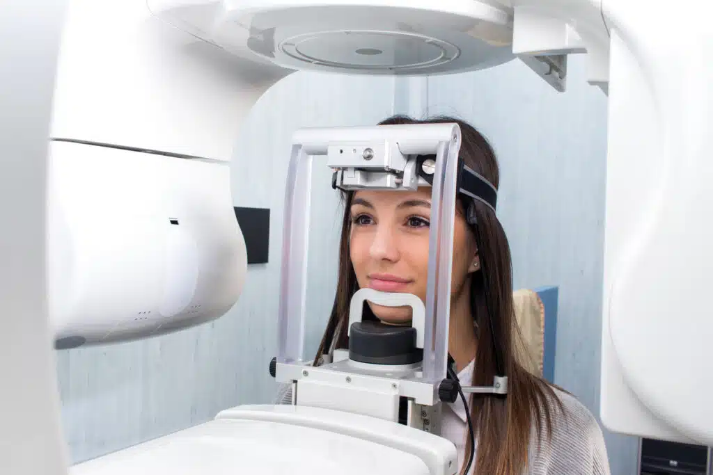
During a Dental CT Scan Procedure
During a dental CT scan, radiologic technologists will position the patient’s head in the machine and then take a series of X-ray images. The patient will be asked to remain as still as possible during the scan and follow any instructions given by the technologist. Once the scan is complete, the patient’s images will be downloaded onto a computer for analysis. This procedure typically takes 15 to 30 minutes, and some patients may experience slight discomfort or nausea during or after the scan due to exposure to radiation.
Additional Information
CT scanners are not currently available in all dentists’ offices. Your dentist may send you to a hospital or radiology center for your exam. If that is the case, it is extremely important that you follow his or her instructions about wearing an appliance during the scan. Dental CT scans are also not covered under most insurance plans, so payment is usually due at the time of the scan.
For more information about dental CT scans and dental implants, contact Gallardo Periodontics and Implant Dentistry today at 305-447-1447.